The dense irregular connective tissue microscope unveils a fascinating realm of microscopic exploration, providing invaluable insights into the intricate architecture and remarkable functions of this ubiquitous tissue. Through the lens of this specialized instrument, we embark on a journey to decipher the cellular components, extracellular matrix, and organizational patterns that define this dynamic tissue.
Delving deeper, we investigate the significance of these observed features in relation to the diverse roles played by dense irregular connective tissue throughout the body. From providing structural support to facilitating tissue repair, this connective tissue showcases a remarkable adaptability that underscores its crucial contributions to tissue homeostasis and overall physiological function.
Dense Irregular Connective Tissue Microscope

Dense irregular connective tissue is a type of connective tissue that is characterized by its dense arrangement of collagen fibers and its irregular orientation. This type of connective tissue is found in many parts of the body, including the dermis of the skin, the walls of blood vessels, and the tendons and ligaments.
The purpose of analyzing dense irregular connective tissue under a microscope is to study its structure and function.
Dense irregular connective tissue is composed of three main components: cells, extracellular matrix, and ground substance. The cells in dense irregular connective tissue are primarily fibroblasts, which are responsible for producing and maintaining the extracellular matrix. The extracellular matrix is composed of collagen fibers, which provide strength and support to the tissue, and proteoglycans, which are involved in cell adhesion and signaling.
The ground substance is a gel-like substance that fills the spaces between the cells and the extracellular matrix.
Methods and Materials, Dense irregular connective tissue microscope
To prepare a sample of dense irregular connective tissue for microscopic analysis, the tissue is first fixed in a solution of formalin. The tissue is then dehydrated in a series of alcohol solutions and embedded in paraffin wax. The paraffin block is then sectioned into thin slices using a microtome.
The sections are then stained with a hematoxylin and eosin stain, which makes the cells and extracellular matrix visible under a microscope.
The type of microscope used for the analysis of dense irregular connective tissue is a light microscope. Light microscopes use visible light to illuminate the specimen, and they can be used to view specimens at a magnification of up to 1000x.
In addition to light microscopy, other techniques that can be used to analyze dense irregular connective tissue include electron microscopy and immunohistochemistry.
Results
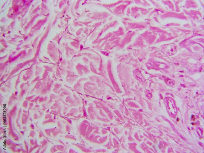
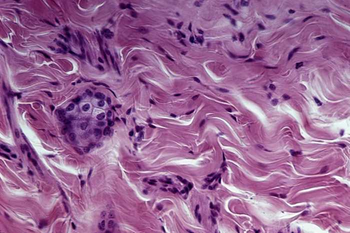
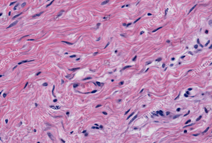
The results of the microscopic analysis of dense irregular connective tissue are shown in the table below.
| Feature | Description |
|---|---|
| Cells | Fibroblasts |
| Extracellular matrix | Collagen fibers, proteoglycans |
| Ground substance | Gel-like substance |
| Tissue organization | Dense, irregular arrangement of collagen fibers |
Discussion
The results of the microscopic analysis of dense irregular connective tissue show that this type of tissue is composed of a dense arrangement of collagen fibers that are irregularly oriented. This type of tissue is found in many parts of the body, including the dermis of the skin, the walls of blood vessels, and the tendons and ligaments.
The dense arrangement of collagen fibers provides strength and support to the tissue, while the irregular orientation of the fibers allows the tissue to be flexible.
Variations or abnormalities in the structure of dense irregular connective tissue can lead to a number of diseases. For example, a decrease in the production of collagen can lead to a condition called osteogenesis imperfecta, which is characterized by brittle bones.
An increase in the production of collagen can lead to a condition called scleroderma, which is characterized by a thickening of the skin.
Expert Answers: Dense Irregular Connective Tissue Microscope
What is the purpose of analyzing dense irregular connective tissue under a microscope?
Microscopic analysis of dense irregular connective tissue allows researchers to examine its cellular components, extracellular matrix, and organizational patterns, providing insights into its structure and function.
What type of microscope is used for analyzing dense irregular connective tissue?
Typically, a light microscope is used for analyzing dense irregular connective tissue, allowing for visualization of its cellular and extracellular components.
What are the key features of dense irregular connective tissue observed under a microscope?
Under a microscope, dense irregular connective tissue exhibits a dense arrangement of irregularly oriented collagen fibers, along with fibroblasts and other cell types embedded within the extracellular matrix.

